Introduction
Tuberculosis is an infectious disease caused by Mycobacterium Tuberculosis, mainly affecting the lungs, but also other parts of the body, known as Extra Pulmonary or EPTB. May be asymptomatic, latent TB or showing active symptoms associated with the organ involved. Immunosuppressive state predisposed to higher incidence of TB. Vocal cord involvement, though rare, may occur in Tb. Symptoms are usually hoarseness of voice.
Case Study
Middle aged, non-smoking, known diabetic patient presented to Outdoor department with complaints of low grade fever since few weeks. CT thorax showed features consistent with sarcoid with elevated Serum Angiotensin Converting Enzyme (ACE) levels. Bronchoscopy guided Transbronchial ling biopsy was normal, with negative BAL culture report.
Patient was started on tapering course of steroids. Clinical symptoms resolved after few months.
Patient was also diagnosed to have ocular sarcoid during his examination for complaints of diminished vision and was put on relevant treatment.
After few months patient complained hoarseness of voice, was referred to ENT surgeon for evaluation. Fiber Optic Laryngoscopy (FOL) showed polypoid lesion in anterior 1/3rd of right vocal cord. Micro laryngeal surgery was performed with excision of right vocal cord polyp and left vocal cord nodule, with samples being sent for Histo Pathological Examination (HPE).
HPE was inconclusive and complaints persisted.
Patient underwent multiple repeat FOL in other setup, was kept on antibiotics, steam inhalation, steroids and other conservative treatment.
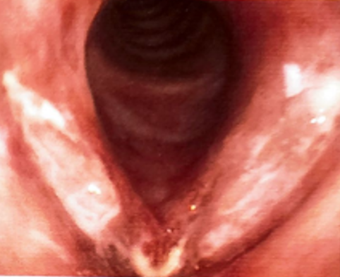
With persisting complaints, another repeat FOL revealed Cobble stoning with dense whitish plaques on both cords and underwent excision biopsy under General Anaesthesia.
HPE showed fragmented tissue bits lined by stratified squamous epithelium and stroma contains collection of histiocytes including some epithelioid morphology favoring epithelioid cell granuloma and areas of necrosis are seen—features suggestive of Koch’s etiology. Gene Xpert detected Rifampicin resistant MTB. The tissue culture came positive for Acud Fast Bacilli Culture on 3rd week and was sent for drug sensitivity testing.
The patient was referred back to Pulmonology Department. Drug Sensitivity Testing(DST) on MGIT-960 showed resistance to isoniazid, pyrazinamide and rifampicin.
Patient was started on all oral longer Multi Drug Resistant Tb regimen including Bedaquilline under Revised National Tuberculosis Control Program (RNTCP).
Patient post discharge reviewed on OPD, with voice much improved and reduced complaints.
Histopathology Report
Specimen—Vocal cord lesion
Gross—Multiple grayish white tissue pieces all together measuring 0.5x0.4x0.3cm-all
Microscopy- Examined sections shows fragmented tissue bits lined by stratified squamous epithelium and stroma contains collections of histiocytes including some of epitheloid morphology favouring epitheloid cell granuloma. Few areas of necrosis are seen.
Interpretation- Chronic necrotizing inflammatory process with occasional ill formed epitheloid granuloma. Consider Kochs etiology.
Discussion
Constitutional symptoms are rare and often hoarseness is the only complaint in laryngeal tuberculosis, which may present even without active pulmonary TB.1, 2 Dysphonia can be caused by — organic (nodules/polyps/tumours/paresis of cords) or functional (functional dysphonia, laryngeal conversion disorder, paradoxical vocal folds motion) or systemic disorders like amyloidosis, sarcoidosis, Wegener’s granulomatosis.3 Mycobacterial infection primarily affected the posterior part of the larynx, which was thought due to pooling of the infected sputum in a recumbent patient. 4 Lymphatic and hematogenous spread associated with an oedematous, polypoid panlaryngitis which is not easily distinguished from chronic laryngitis. 5, 6 Absence of pathognomonic symptoms of laryngeal TB and change in clinical pattern frequently leads to misdiagnosis and delay in the onset of treatment. If untreated, laryngeal TB may lead to widespread local tissue destruction with secondary laryngeal stenosis. 7 Long term steroids for treatment of sarcoidosis, leading to the complete resolution and regression of symptoms, may lead to tuberculosis reactivation or reinfection. 8 Smoking appears to be associated with the development of extensive Laryngeal tuberculosis. 9
Conclusion
Persistent dysphonia should always raise a concern of laryngeal TB particularly in patients on immunosuppressive treatment, even without systemic symptoms. Commonly affected area by this disease are true vocal cord, followed by false vocal cord, epiglottis, arytenoids and posterior commissure. Laryngeal tuberculosis was considered a extra pulmonary manifestation and has almost always been associated with pulmonary infections. Currently reports of isolated laryngeal Tb is on the rise. Appropriate treatment of patients with laryngeal tuberculosis relies on suspicion, prompt diagnosis, and early initiation of appropriate antituberculous treatment.