- Visibility 247 Views
- Downloads 75 Downloads
- Permissions
- DOI 10.18231/j.ijirm.2024.031
-
CrossMark

Mycobacterium abscessus presenting as a lung mass
- Author Details:
-
 Siddharth Ravindranath Waghmare *
-
Snehal Kawade
-
Jairaj P. Nair
Abstract
Introduction: Mycobacterium abscessus is one of the leading causes of non-tuberculous mycobacterial infection. It presents commonly as lung nodules. We report a rare presentation of mycobacterium abscessus as a lung mass.
Case Presentation: A 38-year-old man with complaints of fever, chronic cough, and chest pain presented to our outpatient department. His chest X-ray and CT scan were suggestive of a left lower lobe mass lesion. The sputum cartridge-based nucleic acid amplification test was negative for tuberculosis. The patient was then subjected to bronchoscopy. Mycobacterium abscessus was detected on bronchial washing. This patient was started on a multidrug regime. It consisted of an injection (intramuscular) of amikacin (thrice a week), oral linezolid, clarithromycin, and moxifloxacin. After 1 month of therapy, there was complete resolution of symptoms and X-ray opacity. The patient was continued on the aforementioned treatment.
Conclusion: The clinical presentation of Mycobacterium abscessus is non-specific. In this case report it presented as a mass lesion. This highlights the varied radiological presentation of the infection. Non-tuberculosis mycobacterium infection should be suspected if the initial workup of tuberculosis is negative.
Introduction
Non-tuberculosis mycobacteria (NTM) refers to all the organisms in the genus Mycobacterium except tuberculosis and leprae. Mycobacterium abscessus is a type of rapidly growing NTM. It typically infects those with underlying lung disease. The mode of infection in humans is environmental exposure or undergoing medical or cosmetic procedures. The clinical presentation is indistinguishable from mycobacterium tuberculosis. Due to the high case burden of tuberculosis in India, clinicians rarely consider the diagnosis of NTM. They are often evaluated for tuberculosis and incorrectly treated with anti-tuberculosis treatment. This adds to the morbidity and mortality of the disease. Due to a lack of pathognomonic clinical or radiological features, laboratory diagnosis is the gold standard. A multidrug regime is recommended due to high incidences of drug resistance. The treatment duration is long and response rates are low. We present a case report of mycobacterium abscessus presenting as a lung mass.
Case Presentation
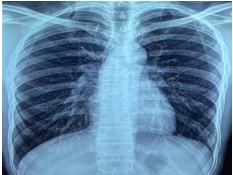
A 38-year-old man presented to the respiratory OPD with complaints of fever, chest pain, and cough. He had these symptoms for 4 months. The patient didn’t suffer from any chronic disease or co-morbidities. He didn’t undergo any clinical procedures. Complete blood count and biochemical investigations were normal. A Chest X-ray demonstrated left lower zone opacity ([Figure 1]). It was confirmed to be an alveolar opacity (without air bronchogram) in the posterior and lateral basal segment of the left lower lobe on computerized tomography of the thorax ([Figure 2]). The sputum cartridge-based nuclei amplification test for negative for tuberculosis. A bronchoscopy was done to collect bronchial wash for culture. Mycobacterium abscessus was isolated via matrix-assisted desorption isonization- time of flight mass spectrometry. The drug sensitivity testing was also performed and the sensitivity pattern is given in [Table 1]. A drug regime consisting of intravenous amikacin, oral linezolid, clarithromycin, and moxifloxacin was started. This regime was designed to follow the drug sensitivity pattern and patient preferences.The patient was reassessed after 1 month of treatment. The symptoms had resolved, and the X-ray opacity had cleared ([Figure 3]).


|
Sensitive |
Intermediate |
Resistant |
|
Ciprofloxacin |
Doxycycline |
Trimethoprim/ Sulfamethoxazole |
|
Moxifloxacin |
Imipenem |
- |
|
Amikacin |
Minocycline |
- |
|
Clarithromycin |
- |
- |
|
Linezolid |
- |
- |
|
Tobramycin |
- |
- |
Discussion
Mycobacterium abscesses is one of the common causes of non-tuberculosis mycobacteria infection encountered in India and the world. It is known to infect skin, soft tissue, and lung disease. M. Abscesses belong to the rapid grower subdivision of the Runyon classification. The risk factors for pulmonary infection are underlying bronchiectasis, cystic fibrosis and a history of tuberculosis. It can only colonize the airways of diseased lungs.[1] Our patient never had tuberculosis or any structural lung disease. Certain professions like gardening and farming are predisposed to the infection due to its presence in the soil and water. Iatrogenic introduction of the bacilli has been observed after medical, dental, and cosmetic procedures. [2], [3] In our case, the patient was a carpenter and hadn’t undergone any such procedures.
The symptoms of the M. abscessus infection depend upon the involved system. Respiratory system involvement presents with symptoms of chronic cough, fever, and chest pain. Often the chronicity of the symptoms is due to the delay in the diagnosis. In our case, the patient had symptoms of cough and chest pain for 4 months. In a TB-endemic country like India, the diagnosis is often delayed due to considerable overlap between the symptomatology of the mycobacterium tuberculosis and abscessus. Chest radiography showed left lower zone opacity in our patient. It was confirmed to be an alveolar mass without an air bronchogram in the left lower lobe on computerized tomography of the thorax. M. Abscessus presents with tree-in-bud appearance (nodules), bronchiectasis, and cavitation. [4], [5] There is no zonal or lobar preponderance. Mediastinal lymphadenopathy is less common. NTM are known to present as lung masses. This type of presentation is commonly seen with mycobacterium avium complex. [6] Due to the non-specificity of the clinical features, microbiological confirmation remains the only method of diagnosis. Sputum, protected bronchoscopic brush sample, or bronchial wash are used to culture the organism in the lab. In accordance with the national tuberculosis elimination programme guidelines, the patient was offered a sputum cartridge-based nucleic acid amplification test. The result was negative for tuberculosis. To confirm the diagnosis, the patient underwent a bronchoscopy. M. Abscessus was isolated by Matrix-assisted desorption isonization- time of flight mass spectrometry from the bronchial wash.
The treatment of lung disease consists of medical management and surgery. M. Abscessus is known to be resistant to macrolide and aminoglycoside.[7] Therefore, it is prudent to perform drug susceptibility testing before treatment. The British Thoracic Society (BTS) guideline recommends sensitivity testing for minimum clarithromycin, cefoxitin, and amikacin. Often antibiotic sensitivity reports aren’t a true representation of in-vivo activity. M abscessus has natural defences against antibiotics like the erm gene. Macrolide resistance is attributed to this gene. It becomes active on prolonged exposure (>14 days) to macrolide antibiotics. [8]
The antibiotics sensitivity pattern in our case is as per Table: 1. The drug regime is based on the clarithromycin sensitivity pattern. As per the BTS recommendations, intravenous medications should be administered for 4 weeks [9] followed by a long term oral regime. In our case, the patient decided against hospitalization or peripherally inserted central catheter insertion line (PICC) insertion. Therefore, the treatment regime was designed as per available the drug susceptibility pattern. The patient was prescribed an injection (intramuscular) of amikacin (thrice a week), and oral linezolid, clarithromycin, and moxifloxacin.
The challenges in medical management are compliance, adverse drug reactions, drug intolerance, long duration of treatment and drug resistance. Myelosuppression and ototoxicity are serious side-effects. [10] A meta-analysis involving 303 patients observed a success rate of 45.6%. [11] After 4 weeks of treatment, there was symptomatic improvement and resolution of X-ray opacity. The treatment will be continued for 1 year beyond culture negativity.
Conclusion
Mycobacterium abscessus often eludes diagnosis due to its nondescript clinical and radiological features. It wasn’t suspected in this case due to a lack of predisposing factors and unusual radiomorphology. Given the high mortality, a high index of suspicious should be exercised and samples(sputum, bronchial wash or tissue samples) should be processed for non-tuberculous mycobacteria.[12] The untoward effects of the drugs and protracted course of antibiotics merit regular follow-up.
Source of Funding
None.
Conflict of Interest
None.
References
- Abdelaal H, Chan E, Young L, Baldwin S, Coler R. Mycobacterium abscessus: It’s Complex. Microorganisms. 2022;10(7). [Google Scholar] [Crossref]
- Gardini G, Gregori N, Matteelli A, Castelli F. Mycobacterial skin infection. Curr Opin Infect Dis. 2022;35(2):79-87. [Google Scholar]
- Chen C, Liu J, Wang S, Yao Y, Yu B, Hu X. Mycobacterium abscessus infection after facial injection of argireline: A case report. World J Clin Cases. 2021;9(8):1996-2000. [Google Scholar]
- Han D, Lee K, Koh W, Yi C, Kim T, Kwon O. Radiographic and CT Findings of Nontuberculous Mycobacterial Pulmonary Infection Caused by Mycobacterium abscessus. AJR Am J Roentgenol. 2003;181(2):513-7. [Google Scholar]
- Chung M, Lee K, Koh W, Lee J, Kim T, Kwon O. Thin-section CT findings of nontuberculous mycobacterial pulmonary diseases: comparison between Mycobacterium avium-intracellulare complex and Mycobacterium abscessus infection. J Korean Med Sci. 2005;20(5):777-83. [Google Scholar]
- Martinez S, Mcadams H, Batchu C. The Many Faces of Pulmonary Nontuberculous Mycobacterial Infection. AJR Am J Roentgenol. 2007;189(1):177-86. [Google Scholar]
- Nessar R, Cambau E, Reyrat JM, Murray A, Gicquel B. Mycobacterium abscessus: a new antibiotic nightmare. J Antimicrob Chemother. 2012;67(4):810-8. [Google Scholar]
- Griffith D, Brown-Elliott B, Benwill J, Wallace R. Mycobacterium abscessus. "Pleased to meet you, hope you guess my name...". Ann Am Thorac Soc. 2015;12(3):436-9. [Google Scholar]
- Haworth C, Banks J, Capstick T, Fisher A, Gorsuch T, Laurenson I. British Thoracic Society Guideline for the management of non-tuberculous mycobacterial pulmonary disease (NTM-PD). BMJ Open Respir Res. 2017;4(1). [Google Scholar] [Crossref]
- Chen J, Zhao L, Mao Y, Ye M, Guo Q, Zhang Y. Clinical Efficacy and Adverse Effects of Antibiotics Used to Treat Mycobacterium abscessus Pulmonary Disease. Front Microbiol. 1977;10. [Google Scholar] [Crossref]
- Kwak N, Dalcolmo MP, Daley CL, Eather G, Gayoso R, Hasegawa N. Mycobacterium abscessus pulmonary disease: individual patient data meta-analysis. Eur Respir J. 2019;54(1). [Google Scholar] [Crossref]
- Jhun B, Moon S, Jeon K, Kwon O, Yoo H, Carriere K. Prognostic factors associated with long-term mortality in 1445 patients with nontuberculous mycobacterial pulmonary disease: a 15-year follow-up study. Eur Respir J. 2020;55(1). [Google Scholar] [Crossref]